Quick version
An ECG, which is both simple and painless, can show how the heart is working by recording its electrical signals. An ECG is used to detect rhythm disorders, lack of oxygen and signs of a heart attack.
Common causes of an ECG are chest pain, palpitations, dizziness or that you belong to a risk group. The doctor interprets the different parts of the curve, such as the P wave, QRS complex and ST segment, to assess the condition of the heart.
A long-term ECG, also called a Holter ECG, records the heart's activity over several days and can detect problems that a regular ECG misses. The earlier heart problems are detected, the better the chances of treatment.
How an ECG works
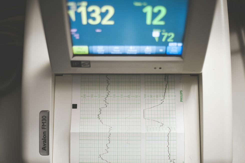
The ECG machine reads the electrical impulses that control the heartbeat. A graph shows the results, with each peak and trough corresponding to a specific part of the heart's work cycle. The curve in the diagram can reveal everything from rhythm disturbances (arrhythmias) to lack of oxygen in the heart muscle.
When should you have an ECG?
If you have symptoms that may indicate a problem with your heart, you should have an ECG, the same applies if you belong to a risk group.
Common reasons for an ECG examination:
Some common reasons for needing an ECG examination are that you:
- experience chest pain or pressure on the chest
- have irregular heartbeats or palpitations
- have dizziness, fainting or experience shortness of breath
- have high blood pressure or a known heart disease in the family
- are about to have surgery or as part of a health check-up
An ECG examination can quickly rule out or confirm suspicions of, for example, heart attack or atrial fibrillation.
Interpreting an ECG - this is what the doctor is looking for
Interpreting an ECG waveform requires both training and experience. To determine how the heart is doing, the doctor looks at different parts of the curve.
Here are some things that are checked:- Rhythm and rate – Is the heart beating regularly and at the right rate?
- P-wave – Indicates how the atria are working.
- QRS complex – Shows how the ventricles are activated.
- ST segment and T-wave – Can reveal oxygen deprivation or heart attack.
- QT interval – A prolonged QT interval can be risky and requires further investigation.
If there are any deviations from normal values, the doctor will receive important clues for further diagnosis.
Frequently asked questions and answers about ECG
Does it hurt to take an ECG?No, an ECG is completely painless. The electrodes are attached to the skin with adhesive strips.
How long does the examination take?The recording itself only takes a few minutes. You will get the results immediately if it is done at a health center or emergency room.
Do I need to prepare?No, but avoid body lotion and oil on your skin before the test - it can impair contact with the electrodes.
Can I do an ECG at home?Yes, there are now portable ECG devices and smart watches that can record simpler ECGs. You can also do a long-term ECG where you wear a small device stuck to your skin for a couple of days. However, these should always be interpreted through a medical assessment by a doctor.
ECG in case of a heart attack - this is what it looks like
In case of a heart attack, blood flow to parts of the heart muscle is blocked - this is something that is clearly visible on an ECG.
Typical signs of an ECG in case of a heart attack:- ST-elevation - A classic sign of an acute infarction.
- T-wave changes - May indicate a lack of oxygen.
- Q-wave formation - Often indicates that the infarction has passed.
An ECG examination is crucial for rapid diagnosis and treatment. A long-term ECG - also called a Holter ECG - records the heart's activity for one or more days. In some cases, it can help to catch temporary signs of oxygen deprivation or rhythm disturbances that a regular resting ECG misses, and thus contribute to the early detection of an incipient heart problem before a heart attack occurs. The earlier a heart attack is detected, the better the chance of recovery.

